
What is Keratoacanthoma(角化棘皮瘤)? - Dr Yeung 楊浩康
Keratoacanthoma is a rapidly growing, locally destructive skin tumor that typically arises from the infundibulum of the hair follicle. Although the precise pathogenesis remains unclear, current research suggests that abnormal regulation of the WNT signaling pathway and mutations in the tumor suppressor gene TP53 may play significant roles in its development. In addition, risk factors such as chronic exposure to ultraviolet (UV) radiation, contact with chemical carcinogens, skin trauma (for example, post-surgical wounds or radiation exposure), and infection with human papillomavirus (HPV) have been implicated in the formation of keratoacanthoma.
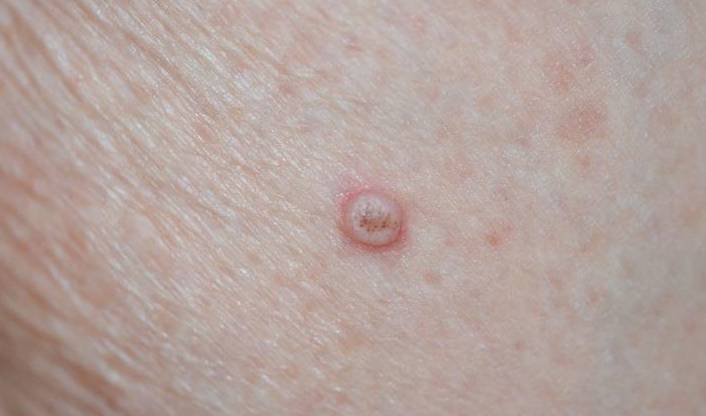
Clinically, keratoacanthoma most frequently manifests as a solitary, round, raised lesion occurring on sun-exposed areas, particularly on the face and upper extremities. The classic appearance is a nodule with a distinctive central crater or depression that is often filled with a keratinous plug, giving it a volcano-like morphology. The surrounding skin is usually smooth and may appear erythematous, sometimes accompanied by mild pruritus or discomfort. Despite its rapid growth, the vast majority of keratoacanthomas are benign; however, their clinical features are often indistinguishable from those of squamous cell carcinoma, basal cell carcinoma, actinic keratosis, or seborrheic keratosis. This similarity necessitates a histopathological examination for a definitive diagnosis.
One of the challenges in managing keratoacanthoma is the common misconception among patients and their families. The term “keratoacanthoma” can evoke fear due to its rapid enlargement and sometimes alarming appearance, leading many to worry excessively about malignancy. In reality, most keratoacanthomas remain benign, though they may occasionally regress spontaneously by forming a crust and eventually healing, often leaving behind a scar. However, because the clinical course is unpredictable and distinguishing it from well-differentiated squamous cell carcinoma can be difficult, surgical excision is frequently recommended to ensure complete removal and to obtain tissue for pathological examination.
For instance, one clinical case involved an elderly patient who presented with multiple keratoacanthomas on sun-damaged skin of the face. The patient was initially very anxious about the possibility of skin cancer. After a biopsy confirmed that the lesions were benign keratoacanthomas, the patient underwent surgical excision combined with cryotherapy. Following treatment, the cosmetic appearance improved significantly and there was no evidence of recurrence during follow-up examinations. Such cases highlight that with proper diagnostic evaluation and timely treatment, keratoacanthoma can be effectively managed and the prognosis is generally excellent.
It is also important to differentiate keratoacanthoma from other skin conditions that may appear similar. Seborrheic keratosis, for example, typically occurs in older adults and presents as a “stuck-on” lesion with a warty, pigmented appearance that lacks the rapid growth seen in keratoacanthoma. Conversely, “malignant freckles” (or dysplastic nevi) display irregular pigmentation and border asymmetry, carrying a potential risk for melanoma. Accurate diagnosis is therefore critical, and physicians rely on clinical examination in conjunction with histopathological analysis to establish the correct diagnosis and rule out malignancy.
In terms of treatment options, various modalities are available based on the size, depth, and number of lesions. For smaller or less aggressive lesions, minimally invasive methods such as cryotherapy (liquid nitrogen treatment), curettage (scraping), or electrocautery (using high-frequency electric current to burn off the lesion) may be sufficient. These procedures not only remove the abnormal tissue but also allow for tissue sampling for further pathological evaluation. In cases where lesions are extensive or exhibit worrying features, surgical excision is the preferred option. This method ensures complete removal of the lesion and allows for precise pathological examination, thereby confirming the diagnosis and excluding a malignancy.
In addition to the direct treatment of keratoacanthoma, preventive measures play a crucial role in long-term management. Patients are encouraged to minimize exposure to UV radiation by wearing protective clothing, using sunscreens with a high SPF, and avoiding prolonged sun exposure, especially during peak hours. Regular skin examinations are recommended for early detection of any new lesions or changes in existing ones. Adopting these preventive strategies can substantially reduce the risk of developing additional precancerous lesions, thereby lowering the potential for malignant transformation.
Furthermore, adjunctive therapies such as topical medications (e.g., 5-fluorouracil or imiquimod) and photodynamic therapy are emerging options that can be used in select cases, particularly for patients with multiple lesions. While these treatments might not be the first line of therapy for keratoacanthoma, they provide additional choices in managing complex cases and may help improve cosmetic outcomes.
In conclusion, keratoacanthoma is a rapidly growing skin tumor with the potential for local destruction, yet it is most often benign. Its development is associated with factors such as UV exposure, genetic mutations, and other environmental influences. Although its appearance may be alarming and similar to malignant skin conditions, proper diagnostic workup—including a biopsy—is essential for accurate differentiation. Treatment strategies range from minimally invasive procedures to complete surgical excision, and preventive measures, including sun protection and regular skin checks, are key to mitigating further risks. With thorough management and appropriate follow-up, patients can achieve excellent outcomes and maintain long-term skin health.